Day 1 :
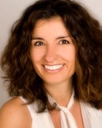
Biography:
She is CEO, Style for your smile, Germany
Abstract:
21st century brought many changes in our lives and the needs of our patients in orthodontics, young or adult, are now much different as they were ten or twenty years ago. Orthodontic treatments all over the world are getting every day faster, more accurate and even more invisible and comfortable as before.
New digital methods such as DVD X-ray s and dental scanns assist orthodontists in creating a perfect smile for every single patient and predict exact treatment results. To describe the treatment outcome with a scan to your patients, it enhances the communication between the doctor and his patients as long as the confidence and the motivation during therapy.
Today´s digital orthodontics allow us to choose from the widest range of treatment options from clear aligners and bracket solution to local lab appliance production. People are looking for stable and good results. Digital technology has helped us to come much more closer to perfection. Now, more than ever, is important that our patients receive the best services. It is what they expect and what they pay for.
Any digital possibility simplifies the everyday clinical workflow for patient diagnosis, treatment planning and model archiving in multiple ways and speeds up your shedule in the practice.
That is where digital orthodontics meet exactly the requirments of a modern Praxis . The majority of our patients are young people. The communicate a lot in the social media and talk about us there. They are proud of a highly compentent and skilled doctor and a high-tech praxis equipment as welle as treatment methods. In the end they recommend us further and contribute to our praxis increasing and developement.
Staying „up to date“ means go digital now! Stay flexible towards your patients and show empathy and compassion to their needs more than ever before, that makes the“ orthodontics NOW“.
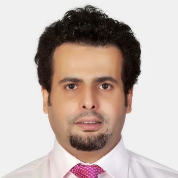
Biography:
Dr. Abdulhakim Alyafei graduated BDD in Damascus University Syria in 1996, in successding years he completed his Postgraduate Diploma in Dental Surgery in 2005; Master Dental Surgery – Pediatric Dentistry in 2007; Advance Diploma in Pediatric Dentisry in 2008 at University of Hongkong. He is a Member of The Royal College of Surgeons of Edinburgh and a Member of The Royal Australian College of Dental Surgeons in Paediatric Dentisry. He also gained his Fellowship in Dental Surgery in The Royal College of Surgeons of Edinburgh England. Dr. Abdulhakim has 20 years of expereince in dentistry with excellent patient’s feedback and exceptional treatment and procedure result. He established new dental department which running all dentistry specialty with advance laboratory and advance dental radiology. He is the first in Qatar to establish the Special Needs Dentistry Clinic along with weekly schedule of Day Care surgery under general anethesia of children. He has involved in 3 publication. Dr. Abdulhakim is currently a Senior Consultant Pedodontist & Special Needs Dentistry; Head of Dental OPD Clinic in Al Wakra Hospital, State of Qatar.
Abstract:
Skeletal and Dental arch anomalies requires intervention at an early age, which may help in avoiding surgical procedures later on and it improves the function of the oral cavity and the facial profile. Inherited and habits as well nasal airways obstruction creates different types of malocclusion. The presentation would demonstrate cases and their treatment plans, supported with updated literature reviews. The following points will be provided;
a. Diagnosis of dental arch anomaly among children;
b. Proper time for intervention;
c. Prevention the complication of arch anomaly;
d. Distinguish between the early need intervention and normal time for orthodontic treatment;
e. Different procedures for early orthodontic treatment.
- Orthodontics| Restorative Dentistry | Dental and Oral Health | Oral and Maxillofacial Surgery
Location: Waterfront 2

Chair
Mridula Goswami
Maulana Azad Institute of Dental Sciences, India
Co-Chair
Andulhakim Alyafei
Al Wakra Hospital, Qatar
Session Introduction
Fahad Mansoor Samadi
King George’s Medical University, India
Title: Association of Candida albicans in development of oral cavity infection with reference to Oral Carcinoma in North Indian population
Biography:
Fahad Mansoor Samadi has completed his MDS from Sardar Patel Post graduate Institute. He is an Assistant Professor in Dept. of Oral Pathology and Microbiology, K G M U. He has published many papers in reputed journals and book chapters.
Abstract:
Presently, cancer one of the most prevalent types of disease is a growing health problem around the world and is one of the leading causes of death. Oral cancer is the sixth most common cancer which occurs worldwide and continues to be the most prevalent cancer which develops in multistep process from pre-existing potentially malignant lesions. The most common precancer is leukoplakia which represents 85% of such lesions and 95% of oral cancers are squamous cell carcinomas (OSCC). In India, the incidence of oral submucous fibrosis (OSMF) and OSCC is also increasing like an epidemic and vast majority of OSCC arises from pre-existing leukoplakia. Several studies have reported that 1-18% of premalignant oral lesions will develop into malignant form. C. albicans has also been identified as a possible factor in development of oral leukoplakia and its malignant transformation. Candida species, dimorphic harmless eukaryotic organism are members of phylum Ascomycota. In healthy individuals, it mostly resides as a part of normal commensal microbial flora on mucosal surfaces of oral cavity. C. albicans grows as a filamentous form, capable of forming true hyphae and is one of the only Candida species. Hyphae play important roles in adhesion and invasion into epithelium. It contributes many virulence attributes like adherence to host tissue and release of some hydrolytic enzymes. It is still unclear, how an increased amount of C. albicans in oral cavity influence the progression of pre-cancer to malignancy. A higher level of C. albicans is present in precancerous and OSCC patients. C. albicans is most pathogenic and significantly more successful pathogen in oral malignancy transformation. There are no drugs which can effect extremely to treat oral cancers. There is a general call for new emerging drugs that are highly effective towards cancer, possess low toxicity, and have a minor environment impact. Novel natural products offer opportunities for innovation in drug discovery. Natural compounds isolated from medicinal plants, as rich sources of novel anticancer drugs, have been of increasing interest. The alarming reports of cancer cases increase the awareness amongst the clinicians and researchers pertaining to investigate newer drug with low toxicity.
Mridula Goswami
Maulana Azad Institute of Dental Sciences, India
Title: Clinical Success In Rediscovering Vitality Through Regenerative Endodontic Therapy

Biography:
Mridula Goswami is Professor & Head, Dept. of Pedodontics & Preventive Dentistry, Maulana Azad Institute of Dental Sciences, New Delhi, India. She has her passion in being with children and expertise in Paediatric Dentistry practice since last 18 years. She is involved in teaching undergraduate and postgraduate courses. Her academic career has many medals & awards. She has to her credit the “Fellowship of International College of Dentists”. Now she is involved in research of her current interest topics which are regenerative endodontics, trauma in children, special need children and lasers in pediatric dentistry.
Abstract:
Statement of the Problem: Pulpal necrosis in an immature tooth with an open apex can have devastating consequences for paediatric dental patients and presents a distinctive challenge for the dentists. Earlier clinicians relied on traditional apexification procedures or the use of apical barriers to treat immature teeth with pulpal necrosis. The latest treatment modalities consist of regeneration-based approaches of tissue engineering.
Theoretical Orientation & Method: Regenerative endodontics is a promising development in the field of tissue engineering involving the diseased or necrotic pulp to be removed and attempted to be replaced with healthy pulp in order to revitalize the teeth. The concept involved in the procedures based on regenerative endodontics is that the platelets in the blood play an important role in hemostasis and wound healing. Dental stem cells and the pulpal connective tissue play a major role in these techniques. Periapical tissues around immature teeth have a rich blood supply and contain stem cells that have relative potential to regenerate in response to tissue injury. This presentation describes two Regenerative Endodontic Techniques i.e. Revascularization procedure and technique using platelet-rich fibrin (PRF). Through clinical case results, it describes the current concepts, treatment approaches and recent developments in the field of ‘Regenerative Endodontics’.
Results: Treatment aims to allow continuation of root development and to regenerate the pulp inside the damaged tooth, preventing the need for routine endodontic treatment. Series of clinical case outcomes show healing of apical periodontitis, promotion of continued root development and restoration of the functional competence of pulpal tissue.
Conclusion & Significance: Continued research, knowledge and understanding can aid in handling scientific challenges of regenerating dental tissues and validating the success of these procedures as an alternative to apexification. With focused research, regeneration of pulp/dentine is likely to be a predictable clinical procedure than a mere vision.
Recent Publications
1.Goswami M, Bhushan U, Goswami M (2016) Dental perspective of rare disease of fanconi anemia: Case report with review. Clin Med Insights Case Rep. 9:25–30.
2. Goswami M, Rajwar AS (205) Evaluation of cavitated and non-cavitated carious lesions using the WHO basic methods, ICDAS-II and laser fluorescence measurements. J Indian Soc Pedod. Prev. Dent. 33(1):10-4.
3.Goswami M, Bhushan U, Mohanty S (2016) Melanotic neuroectodermal tumor of infancy. J Clin Diagn Res. 10(6):ZJ07-8.
4. Singh A, Goswami M, Pradhan G, Han MS, Choi JY, Kapoor S (2015) Cleidocranial dysplasia with normal clavicles: A report of a novel genotype and a review of seven previous cases. Mol Syndromol. 6(2):83-6.
5.Kumar P, Goswami M, Dhillon JK, Rehman F, Thakkar D, Bharti K (2016) Comparative evaluation of microhardness and morphology of permanent tooth enamel surface after laser irradiation and fluoride treatment - An in-vitro study. Laser Therapy 25(3):201-8.
Shaima Nazar
Cairo University, Egypt
Title: Oral Health Related Quality of Life (OHRQoL) among Adults

Biography:
I’m a Kuwaiti dentist, born in Kuwait in 1979. I got my BDS from Cairo University, Egypt in 2001. Since 2001 I’m appointed by the Ministry of Health- Kuwait. In 2002, I worked at the School Oral Health Program as a general pediatric dentist. I participated in a survey in 2004 as an examiner for oral screening of school children. From 2006 to 2016 worked as the Oral Health Education and Prevention team leader. I got MFDS from the RCSI in 2011 and in 2014 Kuwait Board Dental Program Part I. Currently; I’m the Head of Al-Jahra School Oral Health Program.
Abstract:
Objective: The aim of this study was to assess the oral health related quality of life of adults in Kuwait.
Method: A cross- sectional study was done among adults during oral health education activities done by Capital School Oral Program 2012. Self-reported questionnaire was distributed. A convenience sample (N=503) participated in this study. The questionnaire had six sections. One section was about the OHRQoL that consisted of nine questions.
Result: The mean age of participated adults was 35.1±11.08. Females were 43% and 52% were males. Most of participants were healthy. Sixty three percent of participants have college or higher than college level of education. Approximately, quarter of participants (71%) were married with the mean number of children was 3.4±1.6. The OHRQoL measurements showed that 95% of participants enjoyed eating and they liked their smile (80%). Only, 3% had speech difficulties. More than half of participants reported that they never had any social disabilities related to their oral health (62.5%). Most of participants mentioned that they never had any psychosocial disabilities regarding their oral health (79.5%). Overall, 79% of participants judged their oral health as excellent, very good, or good. Seventy nine percent of participants were satisfied about their oral health.
Conclusion: OHRQoL of adults in Kuwait was satisfactory in functional and psychosocial factors related to oral health. Results also indicate that some participants had social disabilities. This can be attributed to high levels of oral diseases among them. National oral health survey among adults should be done to establish this relationship.
Ziyad S. Haid
Profesor Investigador y Director BioMAT'X, Chile
Title: Leukocyte and Platelet-Rich Fibrin in Oro-Maxillo-Facial Surgery: Safety, Efficacy and Clinical Protocol Recommendations from Randomized Controlled Clinical Trials
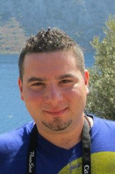
Biography:
Ziyad S. Haidar D.D.S., Cert Implantol, M.Sc. OMFS, MBA, Ph.D. Research Professor and Scientific Director, Faculty of Dentistry, Universidad de los Andes, Santiago de Chile; is a dentist (DDS, AUST, UAE), oral implantologist (Cert Implantol UMDNJ, USA), oral and maxillofacial surgeon (M.Sc., McGill U, CANADA) and Health Care Organization Management Specialist (MBA, JMSB, CANADA) with a doctorate (Ph.D., McGill U, CANADA) in biomaterials, drug delivery and tissue engineering, followed by post-doctoral training at the Shriners Hospital, McGill University Health Center, Montreal, Canada. He is the Founder and Head of the Biomaterials, Pharmaceutical Delivery and Cranio-Maxillo-Facial Tissue Engineering Laboratory and Research Group (BioMAT’X), a newly-established R&D&I unit within the expanding Centro de Investigación Biomédica (CIB) and Facultad de Odontología, Universidad de los Andes in Santiago de Chile. Ziyad is also a Faculty member in the Ph.D.
Abstract:
Objectives: Leukocyte and Platelet-Rich Fibrin (L-PRF) is a 3-D autogenous biomaterial obtained via the simple and rapid centrifugation of whole blood patient samples, in the absence of anti-coagulants, bovine thrombin, additives or any gelifying agents. A relatively new “revolutionary” step in second generation platelet concentrate-based therapeutics, the clinical safety more so, efficacy and effectiveness of L-PRF remains highly-debatable, whether due to preparation protocol variabilities, limited evidence-based scientific and clinical literature and/or inadequate understanding of its bio-components. This critical review provides an update on the application and clinical potential/effectiveness of L-PRF during oral surgery procedures, limited to evidence obtained from human Randomized and Controlled Clinical Trials. Recent functional recommendations on L-PRF preparation protocols are provided to the interested clinician as well as the involved researcher.
Data: All available/accessible clinical trials.
Sources: PubMed (from Jan 2014 – Feb 2016).
Study Selection: Eligibility criteria included: “Human Randomized Controlled Clinical Trials” and “Use of Choukroun’s classic L-PRF preparations only”.
Conclusions: Autologous L-PRF is often associated with early bone formation and maturation; accelerated soft-tissue healing; and reduced post-surgical pain, edema and discomfort. Preparation protocols require revision and standardization. Well-designed RCTs (according to the CONSORT statement) are also needed for validation. Furthermore, a better analysis of rheological properties, bio-components and bioactive function of L-PRF preparations would enhance the cogency, comprehension and therapeutic potential of the reported findings or “observations”; a step closer towards a new era of “super” dental bio-materials and -scaffolds.
Clinical Significance: L-PRF is a simple, malleable and safe biomaterial suitable for use in oral surgery. An innovative tool in Regenerative Dentistry, L-PRF seems a robust and possibly a cost-effective biomaterial alternate for oro-dental tissue repair and regeneration.
Lin-Yang Chi
National Yang-Ming University, Twian
Title: Sugar sweetened beverage is significantly associated with risk of dental caries among school children
Biography:
Lin-Yang Chi completed his PhD at the Cambridge University, UK. He is an Associate Professor at National Yang-Ming University, and the Director of Education and Research Department of Taipei City Hospital, Taiwan. He has published more than 50 papers in reputed journals and has been serving as an Editorial Board Member of Journal of Dental Sciences and other reputated professional journals.
Abstract:
Background & Purpose: According to the results of 2012 national survey in Taiwan, the mean DMFT among 12-year-old school children was 2.50, indicating a significant potential of improvement to be made.
Materials & Method: We used structured questionnaires and standardized oral health check for selected school children to investigate potential risk factors of dental caries, including: caries prevalence, use of fluoride and other prevention measures, caries experience, daily consumption of sugar sweetened food, knowledge and attitude of oral health of main carers, influence of peers, periodic oral health check and self care.
Results: 1856 selected school children of grade 2 and 3 took part in the oral health check, and their parents completed the questionnaires. 965 (51.99%) were boys, mean and SD of age were 8.19 and 0.71 respectively. Those of deft were 4.10±3.21; DMFT 0.90±1.40. The prevalence of caries experience in primary dentition was 82.70%, and permanent dentition 40.84%. Most (1222, 65.84%) of the questionnaires were answered by mothers. 43% of the carers agreed that “water fluoridation is a safe, economic, and effective measure to prevent dental caries”. Similarly, 72% agreed that “fluoride toothpaste is a safe, economic, and effective measure to prevent dental caries”. Multiple regression analysis showed that factors associated with deft of school children were: Gender, age, father’s education, have sugar sweetened drinks, the degree of sweetness of the drinks, and fond of sweets.
Discussion & Suggestions: Our study results showed that most parents had a good understanding for topical fluoridation, and the negative influence of sweets to dental caries. However, only 37% of children used fluoridated toothpaste. There is a gap between what people know and what they do for their children’s oral health. The government, the dental society, and the parents need to take more actions to promote children’s oral health.
Waad Hassan Ibrahim Hassan
University of Science and Technology School of Dentistry, Sudan
Title: Testing Demirjian’s Method of Age Estimation on a Sudanese
Biography:
Waad Hassan Ibrahim Hassan is an Internship Doctor and this research is her graduation research done 2 years ago with her colleagues under the supervision of Dr. Kahlid Mohamed Khalid and with the co-supervisors Wajdi Hassan and Fatima Fath Alrahman.
Abstract:
Introduction: Age estimation is determination of person’s age by using various methods of age estimation for adult and children. The age estimation by using teeth is widely used by many methods and of these methods is the Demirjian method which firstly was introduced in 1973 and used to estimate the age in children.
Aim: The aim of the study was to test the Demirjian method of dental age estimation in Sudanese children population aged (3-16 years).
Methodology: The study design was descriptive retrospective cross sectional hospital based study. The sample was selected using a simple random sampling technique consisting of 500 orthopantograms, (243 boys and 257 girls) collected from Dental Hospital of University of Science and Technology, Esthetica, Almazin and Salah Dafallh’s dental centers. The developmental stages were assessed for each OPG in the left seven mandible permanent teeth and the EA was obtained using Demerjian method and then compared with the chronological age.
Result: The results showed that Demirjian method was not applicable on Sudanese population. It overestimated the age of female samples by about (0.13) years, and under estimated the age of male samples by about (0.46) years. Demirjian method was more reliable in females than males.
Conclusions: Generally Demirjian method was more applicable in females than males and it was more reliable to females in the age group of 15–16 years.
Dheyaa .N. Obada
Baghdad University, College of Dentistry, Iraq
Title: Nano surgical Treatment for Anterior Teeth with Large Periapical Lesion
Biography:
DHEYAA .N.OBADA has graduated from Baghdad university,collage of dentistry at age 23 years starting his researching in 2003 on APEX LOCATER made by him ,in MOROCCOR specialized dental center , in 2005 he made research on bleaching effect and post-operative pain, in DIYALA from 2006 till 2017 he continue his researches on methods and technique that improve the success rate for endodontic treatment with fellow up for 10 years in his private clinic in al-sadder city.
Abstract:
Aim: To investigate sign and symptoms, bactericidal effect for using diode laser 810 nm, and calcium hydroxide, povidone iodine on endodontically treated tooth with periapical abscess
Materials & Methods: Patient with tooth no.9 failed endodontic treatment with periapical abcess, X-ray, diode laser 810 nm, water irrigation, calcium hydroxide, povidone iodine, removing gutta percha from infected tooth, remaining and filling, laser fiber passed over the apical foramen 2 mm, 4 mm according to the size of lesion, lasing 2 sc then stop for 4 sc with water irrigation to avoid over heat, till 20 sc of lasing completed ,apply calcium hydroxide dressing with povidone iodine, examine patient sign and symptom during 8 days, x ray after 6 months.
Result: The patient showed no swelling, sevirity of pain became less, however antibiotic and analgesic are recommended.
Conclusion: using diode laser in PERIAPICAL ABCESS IF WE REACH THIS AREA had less traumatic effect than using a flap ,, less healing time , with cooling technique consideration, and calcium hydroxide and povidone iodine with bactericidal effect on the lateral canal, we will get fine result.
Rajinder Khehra
Cardiff School of Dentistry, UK
Title: An analysis of breakfast cereals marketed to children in the UK with specific regard to oral health.
Biography:
Rajinder Khehra completed her Bachelor of Dental Surgery at the age of 24 from Cardiff School of Dentistry in 2015. Following completion of her dental foundation training in the West Midlands she is now in the middle of her first year in Dental Core Training in which she has completed a 6 month roatation in Oral and Maxillofacial Surgery and currently in a 6 month Community Dental rotation. Her future career aspirations are to become a specialist Orthodontist.
Abstract:
Background
Breakfast cereals remain popular with children and although they are mainly eaten at breakfast time they are regularly eaten between meals because they are quick and easy to prepare. In the UK breakfast cereals are promoted using a wide variety of marketing techniques via a range of media (e.g. TV, radio, magazines, social media) concepts such as the “healthy breakfast option” and a good way to start the day or “fuel your day” predominate. However, many breakfast cereals contain high levels of sugar, some reaching 35% or more. Regular consumption of high sugar breakfast cereals is of concern both from a dental and a general health perspective because of the relationship with dental caries and excess energy intake which could potentially lead to obesity and/or diabetes.
Aim/Objectives: The aim of this study was to ascertain how breakfast cereals in the UK are specifically marketed to children in terms of the nutritional messages portrayed, especially in terms of oral health, both via the written word and visual images.
Methods: 13 of the most popular UK children’s breakfast cereals, including branded and supermarket own-brand versions, were selected for this study. A content analysis was performed using the packaging of each breakfast cereal type; which involved a detailed analysis of the imagery and claims made, together with an assessment of the nutritional content. An additional case study of the most popular brand was undertaken to assess wider media advertising via the internet and social media.
Results: Four of the nine breakfast cereals contained high levels of sugar, according to the UK Traffic Light System which categorises high as in excess of 22.5%; Kellogg’s Frosties contained 37% sugar. The remaining 5 cereals had between 4.4% and 21.4%. With regard to salt and fat, all cereals analysed were labelled as either containing low or medium levels. Supermarket own-brand versions did not differ in nutritional content when compared with the market leader. Nutritional claims focussed on vitamins especially folic acid, iron, whole grains, and no artificial colours or flavours and these were legitimate in terms of nutritional content. Only two opted for the voluntary Front of Pack traffic-light system and these were the supermarket own-brands. A range of marketing techniques were employed, e.g. cartoon characters, royal endorsements, QR codes. The imagery surrounding portion size was grossly misleading.
Conclusion: Some breakfast cereals marketed to children in the UK have very high levels of sugar and the manufacturers use legitimate claims about other nutritional constituents to potentially mislead consumers into thinking the breakfast cereals are healthy. Furthermore, the imagery surrounding portion size is of particular concern when actual portion sizes should be at least a third less than those portrayed on food packaging. Dental and other health professionals need to be aware of the high sugar content of these cereals and the marketing techniques employed by their manufacturers when giving nutritional advice to children and parents.
Saurin Rajnikant Popat
University of Buffalo School of Medicine & Dentistry, USA
Title: Use of Induction Chemotherapy for Advanced Stage Oral Cavity Squamous Cell Carcinoma
Biography:
Saurin R. Popat has completed a Doctorate of Medicne from the University of Western Ontario, London, Canada in 1992, followed by an Otolaryngology – Head & Neck Surgery Residency at the University of Toronto in 1997 and two Fellowships in Head & Neck Surgical Oncology/Microvascular Reconstruction at the University of Toronto (1999) and Skull Base Surgery in Switzerland (2000). He completed an MBA from Cornell University (2010) and serves as Director of Head & Neck Surgery at the Comprehensive Head & Neck Center (CHANCe) in Buffalo, NY with more than 30 publications and book chapters.
Abstract:
Abstrcat:
Oral cavity squamous cell carcinoma (OC-SCCa) accounts for nearly 1/3 of all head and neck squamous cell cancers in the US however with significant international geographic variation due to carcinogen exposure and availability. The mainstay of treatment for resectable disease had been surgery with postoperative radiation based on specific clinical and pathologic features. Survival rates have little improved prior to the use of concurrent postoperative chemoradition. Since 2009, we have employed induction chemotherapy (ICT) as an option to be added to the comprehensive treatment of OC-SCCa. A review of our outcomes was conducted.
Patients/Methods: Retrospective review from 2010 -2015 of patients with stage IV OC-SCCa with minimal 24 month follow-up who received ICT and with no previous history of treated head & neck SCCa. 59 patients were identified however 6 met inclusion criteria. All received 2 to 3 cycles of ICT consisting of cisplatin and either fluorouracil and/or a taxane. Following IHC, all underwent surgical resection.
Results: Survival from OC-SCCa was 100% with 4 patients NED and 2 patients DOC/NED. Median survival was 54.3 months. Three patients had ICT complications but recovered and proceeded to surgery. There were 3 surgical complications in 2 patients. Historic literature based on AJCC data has survial at 4.5 years to be approximately 29%.
Conclusions: Though the study size is small, ICT demonstrated excellent tumor response in our patient population with enduring and superior disease free survival. A larger multicenter trial would be required to confirm these results and outline chemotherapy drug selection.
- Endodontics | Basic Dentistry| Oral and Maxillofacial Surgery| Dental and Oral Health
Location: Waterfront 2
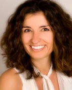
Chair
Eleni Still
, CEO, Style for your smile, Germany
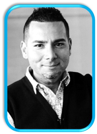
Co-Chair
Ziyad S. Haid
Profesor Investigador y Director BioMAT’X, Chile
Session Introduction
Dheyaa .N. Obada
Baghdad University, College of Dentistry
Title: Emergency Treatment With Diode Laser for Endodontically Treated Tooth With Periapical Abcess Comparing with traditional surgical treatment
Biography:
DHEYAA .N.OBADA has graduated from Baghdad university ,collage of dentistry at age 23 years ,starting his researching in 2003 on APEX LOCATER made by him ,in MOROCCOR specialized dental center ,in 2005 he made research on bleaching effect and post-operative pain, in DIYALA ,from 2006 till 2017 he continue his researches on methods and technique that improve the success rate for endodontic treatment with fellow up for 10 years in his private clinic in al-sader city
Abstract:
Aim: To investigate sign and symptoms, bactericidal effect for using diode laser 810nm ,and calcium hydroxide , povidone iodine on endodontically treated tooth with periapical abscess
Material and Method: Patient with tooth no.9 failed endodontic treatment with periapical abcess,x ray ,diode laser 810nm,water irrigation ,calcium hydroxide ,povidone iodine ,removing gutta percha from infected tooth ,reaming and filling ,laser fibre passed over the apical foramen 2mm ,4mm according to the size of lesion ,,lasing 2sc then stop for 4 sc with water irrigation to avoid over heat ,, till 20 sc of lasing completed ,apply calcium hydroxide dressing with povidone iodine ,examine patient sign and symptom during 8 days ,x ray after 6 months.
Result: The patient shows no swelling, severity of pain became less, however antibiotic and analgesic are recommended.
Conclusion: Using diode laser in PERIAPICAL ABCESS IF WE REACH THIS AREA had less traumatic effect than using a flap ,, less healing time , with cooling technique consideration, and calcium hydroxide and povidone iodine with bactericidal effect on the lateral canal, we will get fine result.
Rajashree Dasari
Panineeya Institute of Dental Sciences and Research Centre, India
Title: Correlation between Expression of Cyp24, Cyp27 and Translocation of VDR in to the Nucleus of Aggressive Periodontitis Patients.
Biography:
Dr.Rajashree Dasari has completed her postgraduation from NTR University of health sciences and currently working as Associate professor at Panineeya Institute of Dental Sciences, India. Her Passion for research and understanding the pathogenesis of periodontal disease creates a new pathways for improving the ability of treating, maintaining the oral health. She also won the best paper award for the research done related to the role of vitamin D receptor at Hongkong International conference, Hongkong in 2014.
Abstract:
Generalized aggressive periodontitis exhibits severe inflammation and alveolar bone loss. 1, 25-dihydroxyvitamin D3 (1, 25(OH) 2D) is the active form of vitamin D3, which plays an important role in calcium and bone metabolism. Vitamin D receptor regulates both bone metabolism and inflammation related genes. It plays a key role in oral homeostasis and its dysfunction may lead to periodontal disease. 1,25(OH)2D activity is regulated by three genes, 25-hydroxyvitamin D-1α-hydroxylase (CYP27), 25-hydroxyvitamin D-24-hydroxylase (CYP24) and vitamin D receptor (VDR). The aim of the present study is to analyze the role of these enzymes in expression of VDR receptor in the nucleus of gingival tissue cells of aggressive periodontitis patients. This study included 31 systemically healthy subjects with aggressive periodontitis and 30 healthy individuals. The level of expression of the above genes was estimated by polymerase chain reaction and the nuclear expression of vitamin D receptor by immunohistochemical analysis. Individuals of both the groups expressed CYP 24 and CYP 27 with medium to low intensity. There was no statistical difference in overall degree of expression of CYP 24 between control and test group. CYP 27 was expressed less in aggressive periodontitis patients than the healthy group [0.461, p>0.01]. VDR expression in the nucleus was more in the healthy patients than the test group. The local tissue specific synthesis of 1,25(OH)2D3 is important as it plays a key role in disease progression. Cyp 24 and Cyp 27 functions in vitamin D target tissues to degrade and generate 1,25(OH)2D3 respectively. Thus the concentration of this enzyme and regulation of its expression is a primary determinant of the overall biological activity within the cells. The factors affecting these enzymes also play an equal role and should be considered. And it can also use as a marker for disease progression.
Young-gun Kim
Yonsei University, Seoul, Republic of Korea
Title: A proposal for safe and efficient injection points of botulinum toxin in temporal region for sleep bruxism
Biography:
Mar 2002 ~ Feb 2008 : DDS, predental course and dentistry, College of Dentistry, Yonsei university, Seoul, Republic of Korea.Mar 2010 ~ Feb 2013 : Residency program, Department of Orofacial pain and Oral medicine, College of Dentistry, Yonsei university, Seoul, Republic of Korea. Apr 2013 ~ Apr 2016 : as a Army surgeon in dental surgery, Republic of Korea Army. May 2016 ~ Present : Fellowship, Department of Orofacial pain and Oral medicine, College of Dentistry, Yonsei university, Seoul, Republic of Korea
Abstract:
The aim of this study was to simplify the optimal temporal areas for safe and reproducible approach for BoNT injections into the temporalis muscle(TM) by carrying out detailed dissections and measurements of the structures in the temporal area, and to virtually represent a topographic mapping of postural relations among the major anatomical structures such as superficial temporal artery, middle temporal vein and temporal branch of the facial nerve in the temporalis muscle of the temporal area.
Nineteen sides of TM from 10 embalmed Korean cadavers were used in this study. The lateral canthus of the eye and tragus were set as landmarks to establish the reference line of this study. The topographies of the superficial temporal artery, middle temporal vein, temporalis tendon, and the temporalis muscle were evaluated. On the disclosed boundary of the muscle, we can visualize an imaginary, rectangular TM in the temporal area. The surface of TM can be divided into 9 equally sized rectangular areas. The topography of studied anatomical structures in these nine compartments was observed and measured from the superficial to deep layers.
After drawing the muscle boundaries, they were divided into nine compartments in order to simplify the relationship between the soft-tissue landmarks and the anatomical structures, and to more facilitate the description of appropriate injection sites. The relative positional ratios between the anatomical structures were constant in all of the specimens. The reference line was first established as C–T, and the distance between C and E' was set as the bottom side of the TM rectangle. The vertical sides of the rectangle were configured at 80% of the bottom side(ratio 5:4). Based on the results of this study, Am, Mu, and Pm were proposed as suitable BoNT injection sites.
